https://youtu.be/6nYyMnAxn_o Cryptococcal Meningoencephalitis explained.
Video: Cryptococcus meningitis explained.
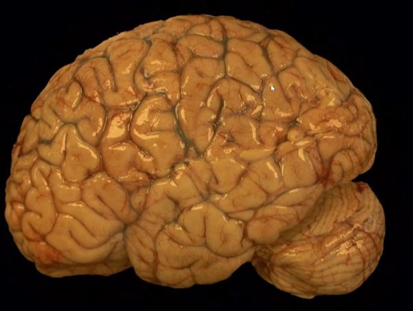
A forum dedicated to the expansion in neuropathology knowledge for students and practitioners alike.
https://youtu.be/6nYyMnAxn_o Cryptococcal Meningoencephalitis explained.


https://youtu.be/qi41D2AgzCg Learn about bacterial infections in the ventricle of a fetus.

A review of the histopathologic diagnosis of the most common primary malignant brain tumor: glioblastoma. https://www.youtube.com/watch?v=3KD6wnMR6Lg&t=74s

https://youtu.be/CHU-464bph8 Review of the histopathologic cancer diagnosis of a carcinoma metastasized to the brain.

https://www.youtube.com/watch?v=87BQ0fuQnqI


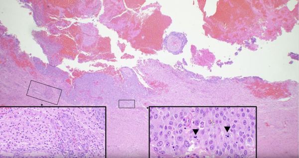
Glioblastomas are high grade astrocytomas that often exhibit microvascular proliferation, characterized by atypical hypertrophic and hyperplastic endothelial cells. A mitotic figure within a proliferating endothelial cell is present in the top right corner of the image.


The massive intraparenchymal hemorrhage depicted in the autopsy specimen of a 60-year-old male patient is the result of hypertensive vasculopathy. Bleeding originated in penetrating vessels of the basal ganglia and extended into adjacent cerebral structures. The blood acts as a space-occupying lesion, resulting in uncal and subfalcine herniation with associated tissue destruction.

")
This middle aged patient had a heterogeneous lesion with multiple irregular rings of enhancement following contrast administration. Biopsy revealed glioblastoma with microvascular proliferation and necrosis, both of which contain leaky blood vessels that contribute to contrast enhancement on imaging.


This elderly patient complaining of headache was diagnosed with glioblastoma following biopsy of the heterogeneous, ring-enhancing lesion in the right temporal lobe. Mass effect caused by the space-occupying tumor has pushed the ipsilateral cingulate gyrus under the falx cerebri, resulting in a subfalcine herniation.