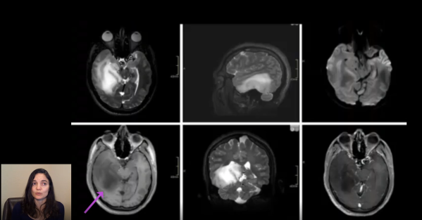
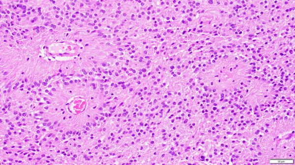
This video is part of a multi-part series reviewing new changes incorporated into the new 5th edition of the WHO classification of tumors of the central nervous system. This video utilizes a case-based format to review important changes in the classification of ependymomas. https://youtu.be/lMVTvFrisbw
Video: Ependymoma – Update from the 5th Edition WHO Classification of CNS Tumors