Malignant Meningitis

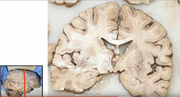
The brain is surrounded by several layers of protective coverings collectively called meninges.  The semi-translucent innermost layers, called the leptomeninges, form a "shrink-wrap" around the brain that allows for easy flow of cerebrospinal fluid (CSF) along the outer surface of central nervous system structures. Unfortunately, it also allows for easy spread of neoplastic cells.  The image shows the inferior aspect... Continue Reading →

Secretory Meningioma: A unique Meningioma variant

The secretory subtype of meningioma (pictured here) is just one of many variants of meningioma, a usually histologically benign tumor that arises from the meninges (i.e. the outer coverings overlying the brain) and often contains concentrically-layered calcifications called psammoma bodies.  The secretory variant can be suspected on imaging by the exuberant edema often seen in the adjacent brain tissue.  Microscopically,... Continue Reading →

Orbital Meningioma with Radiology-Pathology Correlation

Like the brain, the optic nerve is surrounded by meningeal coverings (called the pia, arachnoid and dura) that can infrequently give rise to a meningioma.  Although often histologically benign, intraorbital meningiomas can cause significant morbidity and blindness due to optic nerve compression.  Surgical removal can be curative, as was the case for this patient, whose tumor exhibited the typical... Continue Reading →

Mengioma: Vascular Changes and Prognosis

Meningiomas, like the one pictured here, typically show whorling architectural pattern and nuclear pseduoinclusions.  The vast majority of meningiomas are low grade (WHO Grade 1) dura-based tumors with good prognosis following complete resection.  However, recent studies have suggested that endothelial cell hypertrophy and/or microvascular proliferation in an otherwise typical meningioma are correlated with shorter progression free... Continue Reading →

Website Powered by WordPress.com.

Up ↑

%d bloggers like this: