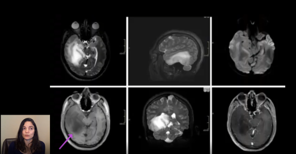
This video is part of a multi-part series reviewing new changes incorporated into the new 5th edition of the WHO classification of tumors of the central nervous system. This video utilizes a case-based format to review important changes in the classification of IDH-mutant and IDH-wildtype diffuse gliomas. https://youtu.be/cfPaeNb04Z8
IDH – Wildtype Astrocytoma: A case-based illustration of updates from the 5th Edition WHO Classification of CNS Tumors
This video is part of a multi-part series reviewing new changes incorporated into the new 5th edition of the WHO classification of tumors of the central nervous system. This video utilizes a case-based format to review important changes in the classification of IDH-mutant and IDH-wildtype diffuse gliomas. https://youtu.be/rskXkeY6DN0
Video: Board Prep – Familial Tumor Syndrome, Mystery Case #3
Mystery case review #3 is one of a series of videos designed to review the variety of brain tumors that arise in the context of a familial tumor syndromes. Test your knowledge with questions posed to the audience in a quiz like format. In-depth answers are provided for each quiz question. This series is perfect... Continue Reading →
Video: Board Prep – Familial Tumor Syndrome, Mystery Case #1
Mystery case review #1 is the first of a series of videos designed to review the variety of brain tumors that arise in the context of a familial tumor syndromes. Test your knowledge with questions posed to the audience in a quiz like format. In-depth answers are provided for each quiz question. This series is... Continue Reading →
Resorption of Embolic Material in Arteriovenous Malformation (AVM)
Vascular brain lesions have increased risk of intracranial bleeding and, therefore, present a challenge to neurosurgeons attempting surgical resection. Such tumors may first be embolized prior to surgical excision in order to reduce the risk of bleeding. Onyx, an ethylene vinyl alcohol copolymer, is one of many embolic agents available to accomplish this task. Onyx has... Continue Reading →
Hippocampal Atrophy
The hippocampus is critical to learning and memory. Patients with hippocampal atrophy often have difficulty with declarative memory (i.e. remembering facts, names, events, etc.) and making new memories. Hippocampal atrophy can be seen a variety of disease processes, including epilepsy and neurodegenerative disease. Note how atrophy in this elderly patient, who had memory difficulties prior to... Continue Reading →
Cerebral Vascular Territories
The cerebral hemispheres are supplied with blood via three major arteries: the anterior, middle, and posterior cerebral arteries. This coronal section through the frontal lobes shows hemorrhage involving the vascular territory of which of these three major cerebral arteries? Answer: The cerebral hemispheres are supplied with blood via three major arteries: the anterior, middle, and posterior... Continue Reading →
Periventricular Leukomalacia (PVL)
Periventricular Leukomalacia (PVL) is a type of brain damage that affects fetuses and premature babies. At this stage of brain development, the white matter surrounding the ventricles is particularly vulnerable to hypoxic (lack of oxygen) or ischemic (lack of blood flow) injury for a variety of reasons, including high metabolic demand in a location that... Continue Reading →
Ice Cream and Imaging: Typical Appearance of Vestibular Schwannoma
Cranial nerve schwannomas most commonly arise from Schwann cells that myelinate the distal aspect of the vestibular division of the 8th cranial nerve. Vestibular schwannomas, sometimes referred to by the double misnomer "acoustic neuroma" (it is a double misnomer because they are not neuromas and they do not usually involve the acoustic division of cranial... Continue Reading →
Classic Imaging: Cyst with an Enhancing Mural Nodule
For many years the only mechanism for observing gross pathologic features of CNS neoplasms was to examine brains extracted after death. However, advancements in imaging technology now allow providers to observe typical gross neuropathological findings in the brains of living patients. Some brain tumors have characteristic MRI findings, an example of which is a cyst... Continue Reading →


