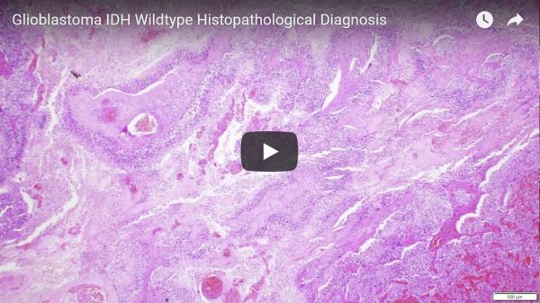
Giant Cell Glioblastoma, also called magnocellular or monstrocellular glioblastoma, is characterized by atypical cells that are extremely large, especially when compared to the non-neoplastic inflammatory cells seen on the left side of the image. The tumor has increased number of dividing cells, including typical mitotic figures (bottom left arrow) and atypical or granular mitoses (bottom... Continue Reading →
Tuberculosis with Caseating Granuloma
Tuberculosis, or TB, is characterized by the formation of granulomas that often have central necrosis, called caseous necrosis due to its cottage-cheese appearance on gross evaluation. Caseating granulomas characteristically have central necrosis surrounded by an immediate layer of macrophages and inflammatory cells, which, in turn, are surrounded by proliferating fibroblasts. This effort to contain the... Continue Reading →
Video: Histopathological diagnosis of Peripheral Neuroblastic Tumors explained (i.e. Neuroblastoma, Ganglioneuroblastoma, and Ganglioneuroma), with an example review of Ganglioneuroma.
Histopathological diagnosis of Peripheral Neuroblastic Tumors explained (i.e. Neuroblastoma, Ganglioneuroblastoma, and Ganglioneuroma), with an example review of Ganglioneuroma. https://youtu.be/w9FEXK8tvJg
Brain trauma and diffuse axon injury in the Corpus Callosum
Diffuse axonal injury: Trauma that is strong enough to break long slender blood vessels is usually strong enough to break long delicate axons, too. Therefore, the presence of gross microhemorrhages in long white matter tracts, such as the corpus callosum, strongly suggests that diffuse axonal injury will be seen on microscopic evaluation of the axons comprising these white matter tracts. The image shows... Continue Reading →
Multiple Sclerosis: Macrophages and Granular Mitosis in MS Plaques
Demyelinating lesions, such as those found in multiple sclerosis, typically contain numerous macrophages with abundant myelin debris filling the cytoplasm. Additionally, atypical and granular mitoses can be seen in demyelinating lesions, though they can also occur in gliomas. Follow us on Facebook, Instagram, and YouTube: Adventures in Neuropathology
Video: Rhabdoid Meningioma explained.
https://youtu.be/YlKgPHM3YzY Follow us on Facebook, Instagram, and YouTube: Adventures in Neuropathology
Video: Glioblastoma IDH Wildtype Histopathological Diagnosis
https://youtu.be/iQ1Be_0IpJM Follow us on Facebook, Instagram, and YouTube: Adventures in Neuropathology
Video: Hemorrhages and hematomas in the Central Nervous system – A narrative of microscopic features.
Hemorrhages and hematomas in the Central Nervous system - A narrative of microscopic features. https://www.youtube.com/watch?v=MCx1DFjANzU&feature=youtu.be Follow us on Facebook, Instagram, and YouTube: Adventures in Neuropathology
Craniopharyngioma on Gross Examination of Brain
This cystic and solid suprasellar mass, seen on this brain cut in the coronal plane, represents the classic gross appearance of a craniopharyngioma with typical focal yellow calcifications. The solid parts on gross examination correlate with enhancing regions on MRI while the cystic regions characteristically contain "machine oil"-like fluid, which is not appreciated in this... Continue Reading →
Video: Acute Traumatic Brain Injury
A review of common gross findings of acute traumatic brain injury (TBI), including a discussion of herniation types and contusional patterns. https://www.youtube.com/watch?v=vnPdJ77khgo&t=333s
