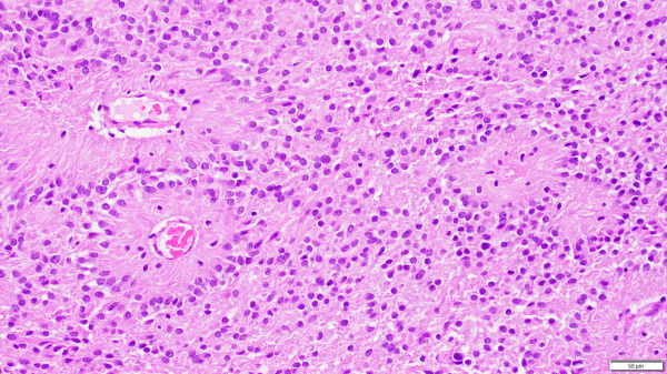
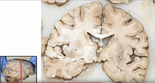
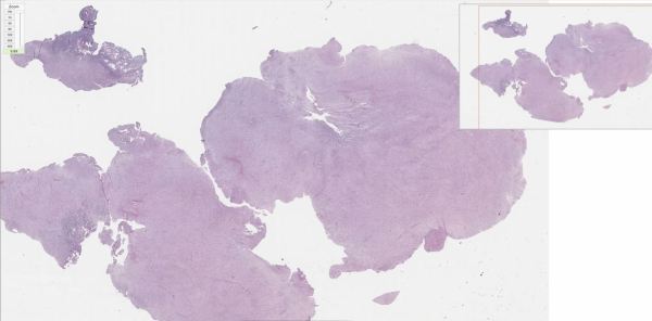
Although meningiomas are classically dura-based lesions, they can also arise in the choroid plexus and, thus, must be considered in the differential diagnosis for intraventricular lesions. This intraventricular meningioma, shown here, is growing underneath normal choroid plexus epithelium.
Video: Ependymomas explained.
Ependymomas are glial tumors that commonly harbor perivascular pseudo-rosettes, seen here, characterized by radially arranged tumor cells around a blood vessel core. https://youtu.be/UXDIYV_yMro
Video: Cryptococcus meningitis explained.
https://youtu.be/6nYyMnAxn_o Cryptococcal Meningoencephalitis explained.
Premature Fetal Brain
The brain of a premature fetus, shown here (front of brain pointing to the left) is initially smooth in the early stages of development. After about 20 weeks gestation, grooves develop in the cortical surface that gradually become more defined until they form well delineated gyri and sulci (i.e. bumps and grooves) typical of a mature brain. The brain... Continue Reading →
Video: Bacterial ventriculitis in prematurity.
https://youtu.be/qi41D2AgzCg Learn about bacterial infections in the ventricle of a fetus.
Video: Glioblastoma Histopathologic Diagnosis
A review of the histopathologic diagnosis of the most common primary malignant brain tumor: glioblastoma. https://www.youtube.com/watch?v=3KD6wnMR6Lg&t=74s
Video: Carcinoma Metastasis to Brain
https://youtu.be/CHU-464bph8 Review of the histopathologic cancer diagnosis of a carcinoma metastasized to the brain.
Video: Rosenthal Fibers in Pilocytic Astrocytoma
https://youtu.be/SzmLY-yh87U Subscribe to our YouTube channel for more educational Neuropathology, and Neuroanatomy videos: https://www.youtube.com/channel/UCw_0moRmVeFNF5kkVHrAU4g/videos?
Ependymoma
A common histologic finding in ependymomas (shown here) are perivascular pseudo-rosettes characterized by neoplastic ependymal cell nuclei radiating outward from a blood vessel, which creates a pink zone of glial processes immediately surrounding the blood vessels.
