Learn about basic histopathologic diagnosis of diffuse astrocytomas. https://www.youtube.com/watch?v=sm764936sIg&feature=share
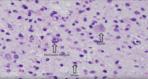
Video: Diffuse Astrocytoma IDH Mutant
A forum dedicated to the expansion in neuropathology knowledge for students and practitioners alike.
Learn about basic histopathologic diagnosis of diffuse astrocytomas. https://www.youtube.com/watch?v=sm764936sIg&feature=share

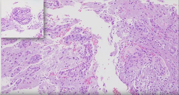
Short segment on recognizing vascular proliferation in Glioblastomas. https://www.youtube.com/watch?v=UefxKiHxwzo


This image of a nerve root shows axons myelinated by oligodendroglial cells of the central nervous system (top of image) and Schwann cells of the peripheral nervous system (bottom of image) at a point of transition called the Obersteiner-Redlich zone.


Coronal section of a fetal brain, approximately 21 weeks gestational age, with edema. Notice how, unlike in adult brains, there is no apparent delineation between white matter and grey matter due to incomplete myelination of the immature brain.


A common histologic finding in ependymomas (shown here) are perivascular pseudo-rosettes characterized by neoplastic ependymal cell nuclei radiating outward from a blood vessel, which creates a pink zone of glial processes immediately surrounding the blood vessels.


Learn about the normal structures, layers and chambers of the human eye. https://www.youtube.com/watch?v=iZCVDtHHa7U&feature=share Subscribe to your Adventures in Neuropathology YouTube channel for more educational neuropathology content: Adventures in Neuropathology

Learn about the histopathologic diagnosis of the secretory variant of meningioma. https://youtu.be/UzUqHRPx1eo Subscribe to your Adventures in Neuropathology YouTube channel for more educational neuropathology content: Adventures in Neuropathology


Stemming from the Latin word "Pilos", meaning "resembling or composed of hair", pilocytic astrocytomas are named as such because of their long hair-like gliofibrillary processes (clear arrows) that stem off of slender bipolar nuclei (black arrows), which are best seen on smear preparation of fresh tissue (depicted here).


Learn about the histopathologic diagnosis of hemangioblastomas, including discussion of other entities that should be considered in the differential. Please subscribe to our YouTube channel for more neuropathology related educational narratives: https://www.youtube.com/channel/UCw_0moRmVeFNF5kkVHrAU4g/featured https://www.youtube.com/watch?v=NozhN6Qk5yo


The Human eye is divided into two main regions: 1. the vitreous humor-filled vitreous body (bottom of image), and 2. the anterior chamber (top of image), which is bordered by the cornea anteriorly (very top of image) and the iris & lens posteriorly (mid-horizon of image). The disc-shaped lens is typically optically clear to allow... Continue Reading →

