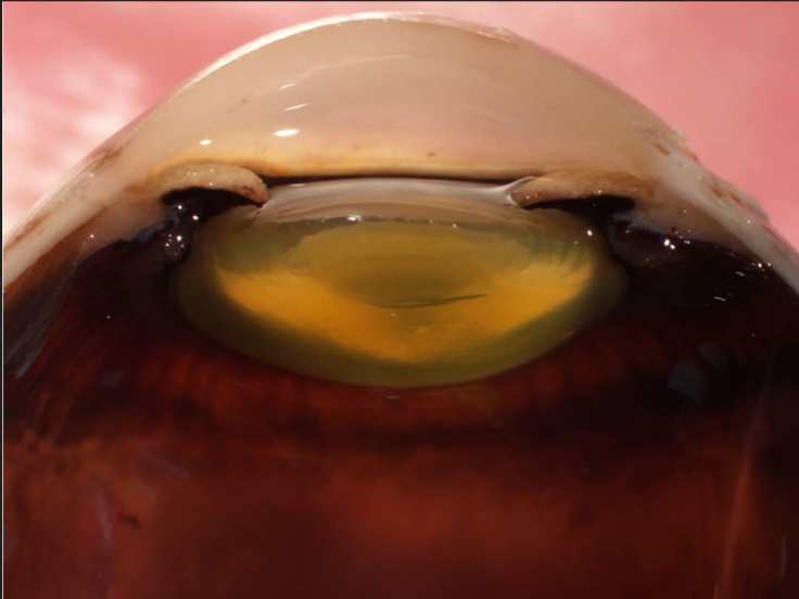
The Human eye is divided into two main regions:
1. the vitreous humor-filled vitreous body (bottom of image), and 2. the anterior chamber (top of image), which is bordered by the cornea anteriorly (very top of image) and the iris & lens posteriorly (mid-horizon of image). The disc-shaped lens is typically optically clear to allow the passage of light to the retina, but it will take on an opaque yellow color after formalin fixation.
.
.
Subscribe to the Adventures in Neuropathology YouTube channel for narrated educational segments: https://www.youtube.com/channel/UCw_0moRmVeFNF5kkVHrAU4g


Leave a comment