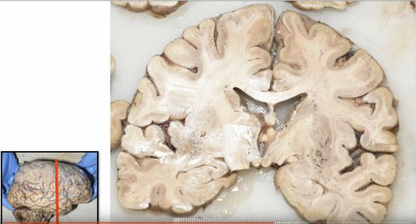
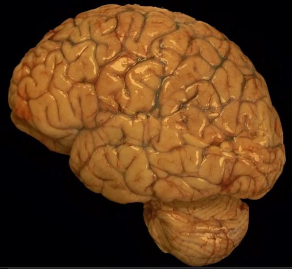
Cerebral edema, or increased swelling in the brain, can occur as the result of numerous etiologies, such as infection, inflammation, metabolic derangement, or neoplastic processes. Typically the brain has an undulating contour featuring crests or bumps called gyri, and troughs or grooves, called sulci. The mass effect produced by cerebral edema results in pushing or... Continue Reading →
Brain trauma and diffuse axon injury in the Corpus Callosum
Diffuse axonal injury: Trauma that is strong enough to break long slender blood vessels is usually strong enough to break long delicate axons, too. Therefore, the presence of gross microhemorrhages in long white matter tracts, such as the corpus callosum, strongly suggests that diffuse axonal injury will be seen on microscopic evaluation of the axons comprising these white matter tracts. The image shows... Continue Reading →
Renal Cell Carcinoma Embolized with PVA
Renal cell carcinoma, a relatively common cancer of the kidney, is a highly vascular lesion that will typically bleed extensively during surgery. Just prior to surgery this renal cell carcinoma that had metastasized to the paraspinal soft tissue was embolized using PVA (polyvinyl alcohol), the blue foreign embolic material within the vessel lumen. This process of embolization was... Continue Reading →
Video: Cryptococcus meningitis explained.
https://youtu.be/6nYyMnAxn_o Cryptococcal Meningoencephalitis explained.
Premature Fetal Brain
The brain of a premature fetus, shown here (front of brain pointing to the left) is initially smooth in the early stages of development. After about 20 weeks gestation, grooves develop in the cortical surface that gradually become more defined until they form well delineated gyri and sulci (i.e. bumps and grooves) typical of a mature brain. The brain... Continue Reading →
Video: Bacterial ventriculitis in prematurity.
https://youtu.be/qi41D2AgzCg Learn about bacterial infections in the ventricle of a fetus.
Video: Glioblastoma Histopathologic Diagnosis
A review of the histopathologic diagnosis of the most common primary malignant brain tumor: glioblastoma. https://www.youtube.com/watch?v=3KD6wnMR6Lg&t=74s
Video: Rosenthal Fibers in Pilocytic Astrocytoma
https://youtu.be/SzmLY-yh87U Subscribe to our YouTube channel for more educational Neuropathology, and Neuroanatomy videos: https://www.youtube.com/channel/UCw_0moRmVeFNF5kkVHrAU4g/videos?
Ependymoma
A common histologic finding in ependymomas (shown here) are perivascular pseudo-rosettes characterized by neoplastic ependymal cell nuclei radiating outward from a blood vessel, which creates a pink zone of glial processes immediately surrounding the blood vessels.