Ice Cream and Imaging: Typical Appearance of Vestibular Schwannoma

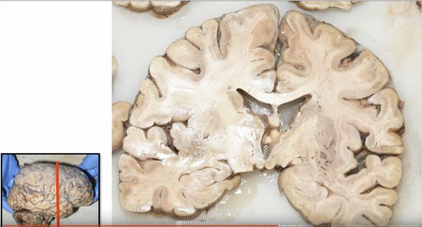
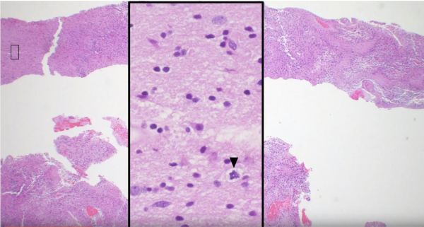
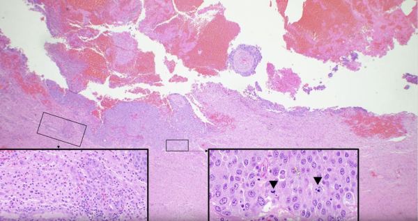
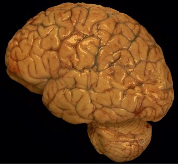
Cranial nerve schwannomas most commonly arise from Schwann cells that myelinate the distal aspect of the vestibular division of the 8th cranial nerve. Vestibular schwannomas, sometimes referred to by the double misnomer "acoustic neuroma" (it is a double misnomer because they are not neuromas and they do not usually involve the acoustic division of cranial... Continue Reading →

Brain trauma and diffuse axon injury in the Corpus Callosum

Diffuse axonal injury: Trauma that is strong enough to break long slender blood vessels is usually strong enough to break long delicate axons, too.  Therefore, the presence of gross microhemorrhages in long white matter tracts, such as the corpus callosum, strongly suggests that diffuse axonal injury will be seen on microscopic evaluation of the axons comprising these white matter tracts. The image shows... Continue Reading →

Renal Cell Carcinoma Embolized with PVA

Renal cell carcinoma, a relatively common cancer of the kidney, is a highly vascular lesion that will typically bleed extensively during surgery.  Just prior to surgery this  renal cell carcinoma that had metastasized to the paraspinal soft tissue was embolized using PVA (polyvinyl alcohol), the blue foreign embolic material within the vessel lumen.  This process of embolization was... Continue Reading →

Premature Fetal Brain

The brain of a premature fetus, shown here (front of brain pointing to the left) is initially smooth in the early stages of development.  After about 20 weeks gestation, grooves develop in the cortical surface that gradually become more defined until they form well delineated gyri and sulci (i.e. bumps and grooves) typical of a mature brain.  The brain... Continue Reading →

Website Powered by WordPress.com.

Up ↑

%d bloggers like this: