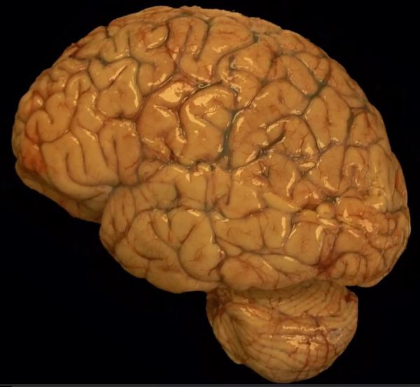
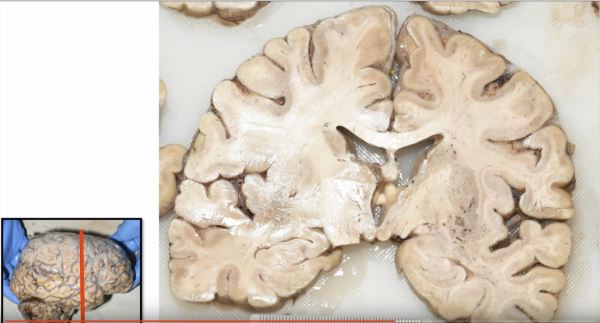
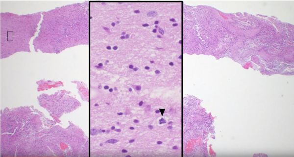
https://youtu.be/6nYyMnAxn_o Cryptococcal Meningoencephalitis explained.
Video: Cryptococcus meningitis explained.
A forum dedicated to the expansion in neuropathology knowledge for students and practitioners alike.
https://youtu.be/6nYyMnAxn_o Cryptococcal Meningoencephalitis explained.


https://youtu.be/qi41D2AgzCg Learn about bacterial infections in the ventricle of a fetus.

A review of the histopathologic diagnosis of the most common primary malignant brain tumor: glioblastoma. https://www.youtube.com/watch?v=3KD6wnMR6Lg&t=74s

https://youtu.be/SzmLY-yh87U Subscribe to our YouTube channel for more educational Neuropathology, and Neuroanatomy videos: https://www.youtube.com/channel/UCw_0moRmVeFNF5kkVHrAU4g/videos?


A common histologic finding in ependymomas (shown here) are perivascular pseudo-rosettes characterized by neoplastic ependymal cell nuclei radiating outward from a blood vessel, which creates a pink zone of glial processes immediately surrounding the blood vessels.


Hemangioblastoma is highly vascular tumor with neoplastic stromal/interstitial cells that have a variably clear cell appearance due to their lipid and glycogen cytoplasmic contents. Hemangioblastomas can be found in patients with von Hippel Lindau (VHL) syndrome, who also have increased risk of developing renal cell carcinoma (RCC). Immunostains can be used to differentiate the inhibin-positive... Continue Reading →


Choroid plexus papillomas are low grade tumors that arise from the intraventricular CSF-producing choroid plexus. Here we see the transition from the round bland nuclei and ample pink cytoplasm of the normal choroid plexus epithelium (bottom of image) to the dysplastic columnar epithelium of the papilloma (top of image) featuring nuclear crowding and mitotic activity... Continue Reading →


Dorsal root ganglia are located along the length of the spinal cord and are composed of clusters of large neuron cell bodies, each with a prominent nucleus and nucleolus, that belong to sensory nerves whose axons deliver sensory information to the spinal cord.