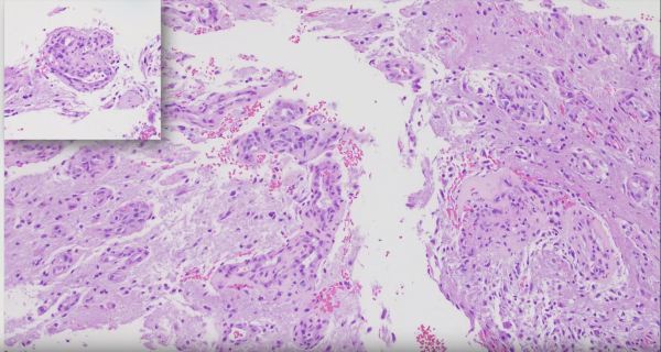
Vascular brain lesions have increased risk of intracranial bleeding and, therefore, present a challenge to neurosurgeons attempting surgical resection. Such tumors may first be embolized prior to surgical excision in order to reduce the risk of bleeding. Onyx, an ethylene vinyl alcohol copolymer, is one of many embolic agents available to accomplish this task. Onyx has... Continue Reading →
Cerebral Vascular Territories
The cerebral hemispheres are supplied with blood via three major arteries: the anterior, middle, and posterior cerebral arteries. This coronal section through the frontal lobes shows hemorrhage involving the vascular territory of which of these three major cerebral arteries? Answer: The cerebral hemispheres are supplied with blood via three major arteries: the anterior, middle, and posterior... Continue Reading →
Arteriovenous Malformation
Arteriovenous malformation, or AVM, is a collection of abnormal vessels that lack an intervening capillary bed and are separated by gliotic brain tissue. Some vessels have thick walls with elastic laminae typical of arteries, while others have thin collagenous walls typical of veins. In addition, "arterialization of a vein" can also be seen in which... Continue Reading →
Intraparenchymal Hemorrhage
The massive intraparenchymal hemorrhage depicted in the autopsy specimen of a 60-year-old male patient is the result of hypertensive vasculopathy. Bleeding originated in penetrating vessels of the basal ganglia and extended into adjacent cerebral structures. The blood acts as a space-occupying lesion, resulting in uncal and subfalcine herniation with associated tissue destruction.
Video: Recognizing Vascular Proliferation in Glioblastomas
Short segment on recognizing vascular proliferation in Glioblastomas. https://www.youtube.com/watch?v=UefxKiHxwzo