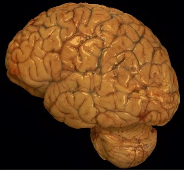
The brain of a premature fetus, shown here (front of brain pointing to the left) is initially smooth in the early stages of development. After about 20 weeks gestation, grooves develop in the cortical surface that gradually become more defined until they form well delineated gyri and sulci (i.e. bumps and grooves) typical of a mature brain. The brain... Continue Reading →
Video: Glioblastoma Histopathologic Diagnosis
A review of the histopathologic diagnosis of the most common primary malignant brain tumor: glioblastoma. https://www.youtube.com/watch?v=3KD6wnMR6Lg&t=74s
Video: Carcinoma Metastasis to Brain
https://youtu.be/CHU-464bph8 Review of the histopathologic cancer diagnosis of a carcinoma metastasized to the brain.
Video: Rosenthal Fibers in Pilocytic Astrocytoma
https://youtu.be/SzmLY-yh87U Subscribe to our YouTube channel for more educational Neuropathology, and Neuroanatomy videos: https://www.youtube.com/channel/UCw_0moRmVeFNF5kkVHrAU4g/videos?
Glioblastomas
Glioblastomas are high grade astrocytomas that often exhibit microvascular proliferation, characterized by atypical hypertrophic and hyperplastic endothelial cells. A mitotic figure within a proliferating endothelial cell is present in the top right corner of the image.
Glioblastoma with Ring Enhancement (MRI)
This middle aged patient had a heterogeneous lesion with multiple irregular rings of enhancement following contrast administration. Biopsy revealed glioblastoma with microvascular proliferation and necrosis, both of which contain leaky blood vessels that contribute to contrast enhancement on imaging.
MRI of Glioblastoma with Subfalcine Herniation
This elderly patient complaining of headache was diagnosed with glioblastoma following biopsy of the heterogeneous, ring-enhancing lesion in the right temporal lobe. Mass effect caused by the space-occupying tumor has pushed the ipsilateral cingulate gyrus under the falx cerebri, resulting in a subfalcine herniation.
Glioblastoma with Pseudopalisading Necrosis
Glioblastomas are malignant astrocytomas that often show pseudopalisading necrosis, characterized by palisading of neoplastic cells along the edges of tumor necrosis. Gioblastomas are the most common malignant primary brain tumor.
Video: Diffuse Astrocytoma IDH Mutant
Learn about basic histopathologic diagnosis of diffuse astrocytomas. https://www.youtube.com/watch?v=sm764936sIg&feature=share
