Short segment on recognizing vascular proliferation in Glioblastomas. https://www.youtube.com/watch?v=UefxKiHxwzo
Video: Recognizing Vascular Proliferation in Glioblastomas
A forum dedicated to the expansion in neuropathology knowledge for students and practitioners alike.
Short segment on recognizing vascular proliferation in Glioblastomas. https://www.youtube.com/watch?v=UefxKiHxwzo


A common histologic finding in ependymomas (shown here) are perivascular pseudo-rosettes characterized by neoplastic ependymal cell nuclei radiating outward from a blood vessel, which creates a pink zone of glial processes immediately surrounding the blood vessels.


Stemming from the Latin word "Pilos", meaning "resembling or composed of hair", pilocytic astrocytomas are named as such because of their long hair-like gliofibrillary processes (clear arrows) that stem off of slender bipolar nuclei (black arrows), which are best seen on smear preparation of fresh tissue (depicted here).


Hemangioblastoma is highly vascular tumor with neoplastic stromal/interstitial cells that have a variably clear cell appearance due to their lipid and glycogen cytoplasmic contents. Hemangioblastomas can be found in patients with von Hippel Lindau (VHL) syndrome, who also have increased risk of developing renal cell carcinoma (RCC). Immunostains can be used to differentiate the inhibin-positive... Continue Reading →


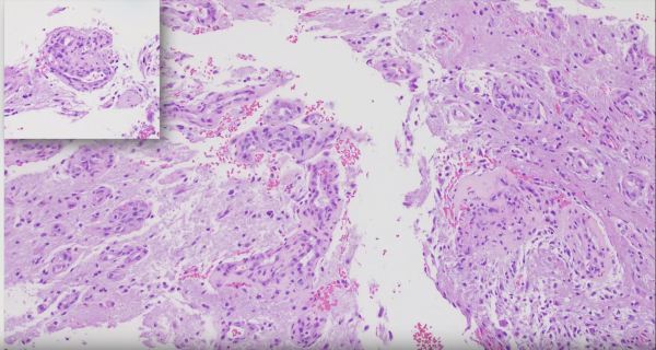
Choroid plexus papillomas are low grade tumors that arise from the intraventricular CSF-producing choroid plexus. Here we see the transition from the round bland nuclei and ample pink cytoplasm of the normal choroid plexus epithelium (bottom of image) to the dysplastic columnar epithelium of the papilloma (top of image) featuring nuclear crowding and mitotic activity... Continue Reading →

