A review of the histopathologic diagnosis of the most common primary malignant brain tumor: glioblastoma. https://www.youtube.com/watch?v=3KD6wnMR6Lg&t=74s
Video: Glioblastoma Histopathologic Diagnosis
A forum dedicated to the expansion in neuropathology knowledge for students and practitioners alike.
A review of the histopathologic diagnosis of the most common primary malignant brain tumor: glioblastoma. https://www.youtube.com/watch?v=3KD6wnMR6Lg&t=74s


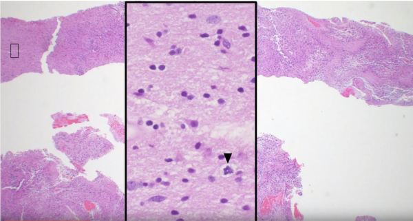
Glioblastomas are high grade astrocytomas that often exhibit microvascular proliferation, characterized by atypical hypertrophic and hyperplastic endothelial cells. A mitotic figure within a proliferating endothelial cell is present in the top right corner of the image.

")
This middle aged patient had a heterogeneous lesion with multiple irregular rings of enhancement following contrast administration. Biopsy revealed glioblastoma with microvascular proliferation and necrosis, both of which contain leaky blood vessels that contribute to contrast enhancement on imaging.


This elderly patient complaining of headache was diagnosed with glioblastoma following biopsy of the heterogeneous, ring-enhancing lesion in the right temporal lobe. Mass effect caused by the space-occupying tumor has pushed the ipsilateral cingulate gyrus under the falx cerebri, resulting in a subfalcine herniation.

Short segment on recognizing vascular proliferation in Glioblastomas. https://www.youtube.com/watch?v=UefxKiHxwzo


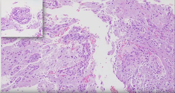
A common histologic finding in ependymomas (shown here) are perivascular pseudo-rosettes characterized by neoplastic ependymal cell nuclei radiating outward from a blood vessel, which creates a pink zone of glial processes immediately surrounding the blood vessels.

