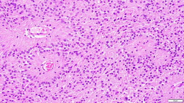
Ependymomas are glial tumors that commonly harbor perivascular pseudo-rosettes, seen here, characterized by radially arranged tumor cells around a blood vessel core. https://youtu.be/UXDIYV_yMro
Video: Ependymomas explained.
A forum dedicated to the expansion in neuropathology knowledge for students and practitioners alike.
Ependymomas are glial tumors that commonly harbor perivascular pseudo-rosettes, seen here, characterized by radially arranged tumor cells around a blood vessel core. https://youtu.be/UXDIYV_yMro

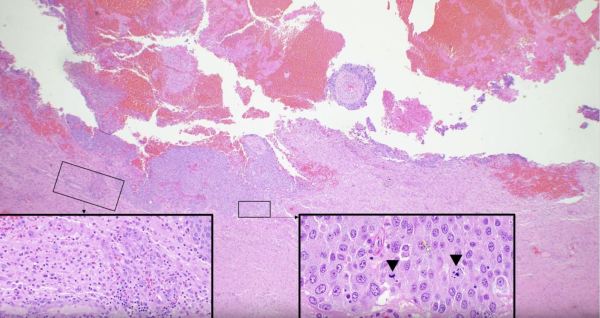
https://youtu.be/6nYyMnAxn_o Cryptococcal Meningoencephalitis explained.


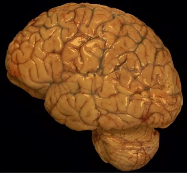
The brain of a premature fetus, shown here (front of brain pointing to the left) is initially smooth in the early stages of development. After about 20 weeks gestation, grooves develop in the cortical surface that gradually become more defined until they form well delineated gyri and sulci (i.e. bumps and grooves) typical of a mature brain. The brain... Continue Reading →


https://youtu.be/qi41D2AgzCg Learn about bacterial infections in the ventricle of a fetus.

A review of the histopathologic diagnosis of the most common primary malignant brain tumor: glioblastoma. https://www.youtube.com/watch?v=3KD6wnMR6Lg&t=74s

https://youtu.be/CHU-464bph8 Review of the histopathologic cancer diagnosis of a carcinoma metastasized to the brain.

https://youtu.be/SzmLY-yh87U Subscribe to our YouTube channel for more educational Neuropathology, and Neuroanatomy videos: https://www.youtube.com/channel/UCw_0moRmVeFNF5kkVHrAU4g/videos?


The World Health Organization Classification of Tumors of the CNS officially recognizes 13 different variants of meningioma, most of which are Grade 1 tumors that are potentially curable with complete resection. Chordoid meningioma is a rare subtype that accounts for less than 1% of all intracranial meningiomas. They are commonly composed of epithelioid tumor cells,... Continue Reading →