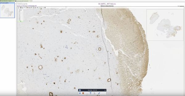
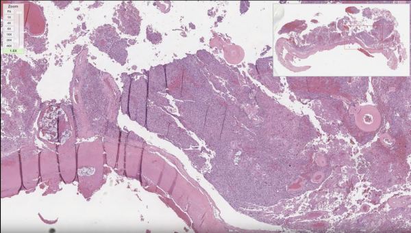
Learn about the histopathologic diagnosis of hemangioblastomas, including discussion of other entities that should be considered in the differential. Please subscribe to our YouTube channel for more neuropathology related educational narratives: https://www.youtube.com/channel/UCw_0moRmVeFNF5kkVHrAU4g/featured https://www.youtube.com/watch?v=NozhN6Qk5yo
Anatomy of the Human Eye
The Human eye is divided into two main regions: 1. the vitreous humor-filled vitreous body (bottom of image), and 2. the anterior chamber (top of image), which is bordered by the cornea anteriorly (very top of image) and the iris & lens posteriorly (mid-horizon of image). The disc-shaped lens is typically optically clear to allow... Continue Reading →
Hemangioblastoma
Hemangioblastoma is highly vascular tumor with neoplastic stromal/interstitial cells that have a variably clear cell appearance due to their lipid and glycogen cytoplasmic contents. Hemangioblastomas can be found in patients with von Hippel Lindau (VHL) syndrome, who also have increased risk of developing renal cell carcinoma (RCC). Immunostains can be used to differentiate the inhibin-positive... Continue Reading →
Video: Cerebral Amyloid Angiopathy
Review of brain labeled with amyloid immunostain that reveals Cerebral Amyloid Angiopathy and Alzheimer-type amyloid plaques. https://www.youtube.com/watch?v=B7UKd-aSuxQ Subscribe to our YouTube channel for more Neuropathology educational topics: https://www.youtube.com/channel/UCw_0moRmVeFNF5kkVHrAU4g
Video: Embolized Meningioma
Basic histopathologic diagnosis of a meningioma that has been embolized. https://www.youtube.com/watch?v=4B0P5xwvsFM
Choroid Plexus Papilloma
Choroid plexus papillomas are low grade tumors that arise from the intraventricular CSF-producing choroid plexus. Here we see the transition from the round bland nuclei and ample pink cytoplasm of the normal choroid plexus epithelium (bottom of image) to the dysplastic columnar epithelium of the papilloma (top of image) featuring nuclear crowding and mitotic activity... Continue Reading →
Dorsal Root Ganglia
Dorsal root ganglia are located along the length of the spinal cord and are composed of clusters of large neuron cell bodies, each with a prominent nucleus and nucleolus, that belong to sensory nerves whose axons deliver sensory information to the spinal cord.
Astrocytes and the Blood Brain Barrier
Astrocytes, like the one highlighted here by GFAP stain, are star-shaped glial cells that are involved in a large number of CNS functions, including the blood brain barrier. Together with pericytes and endothelial cells, astrocyte end plates surrounding a vessel forms an essential component of the blood brain barrier which severely limits passage of certain... Continue Reading →
