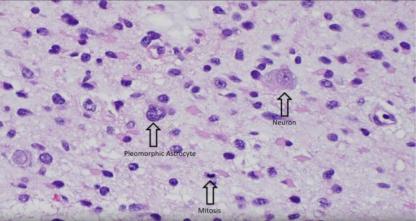
The massive intraparenchymal hemorrhage depicted in the autopsy specimen of a 60-year-old male patient is the result of hypertensive vasculopathy. Bleeding originated in penetrating vessels of the basal ganglia and extended into adjacent cerebral structures. The blood acts as a space-occupying lesion, resulting in uncal and subfalcine herniation with associated tissue destruction.
Intraparenchymal Hemorrhage


")