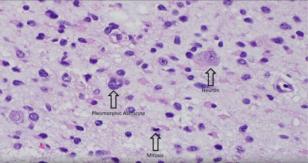
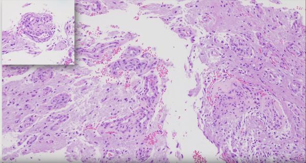
The World Health Organization Classification of Tumors of the CNS officially recognizes 13 different variants of meningioma, most of which are Grade 1 tumors that are potentially curable with complete resection. Chordoid meningioma is a rare subtype that accounts for less than 1% of all intracranial meningiomas. They are commonly composed of epithelioid tumor cells,... Continue Reading →
Glioblastoma with Pseudopalisading Necrosis
Glioblastomas are malignant astrocytomas that often show pseudopalisading necrosis, characterized by palisading of neoplastic cells along the edges of tumor necrosis. Gioblastomas are the most common malignant primary brain tumor.
Video: Diffuse Astrocytoma IDH Mutant
Learn about basic histopathologic diagnosis of diffuse astrocytomas. https://www.youtube.com/watch?v=sm764936sIg&feature=share
Video: Recognizing Vascular Proliferation in Glioblastomas
Short segment on recognizing vascular proliferation in Glioblastomas. https://www.youtube.com/watch?v=UefxKiHxwzo
Coronal Section of a Fetal Brain
Coronal section of a fetal brain, approximately 21 weeks gestational age, with edema. Notice how, unlike in adult brains, there is no apparent delineation between white matter and grey matter due to incomplete myelination of the immature brain.
Ependymoma
A common histologic finding in ependymomas (shown here) are perivascular pseudo-rosettes characterized by neoplastic ependymal cell nuclei radiating outward from a blood vessel, which creates a pink zone of glial processes immediately surrounding the blood vessels.Â
Pilocytic Astrocytoma
Stemming from the Latin word "Pilos", meaning "resembling or composed of hair", pilocytic astrocytomas are named as such because of their long hair-like gliofibrillary processes (clear arrows) that stem off of slender bipolar nuclei (black arrows), which are best seen on smear preparation of fresh tissue (depicted here).
Hemangioblastoma
Hemangioblastoma is highly vascular tumor with neoplastic stromal/interstitial cells that have a variably clear cell appearance due to their lipid and glycogen cytoplasmic contents. Hemangioblastomas can be found in patients with von Hippel Lindau (VHL) syndrome, who also have increased risk of developing renal cell carcinoma (RCC). Immunostains can be used to differentiate the inhibin-positive... Continue Reading →
Choroid Plexus Papilloma
Choroid plexus papillomas are low grade tumors that arise from the intraventricular CSF-producing choroid plexus. Here we see the transition from the round bland nuclei and ample pink cytoplasm of the normal choroid plexus epithelium (bottom of image) to the dysplastic columnar epithelium of the papilloma (top of image) featuring nuclear crowding and mitotic activity... Continue Reading →
