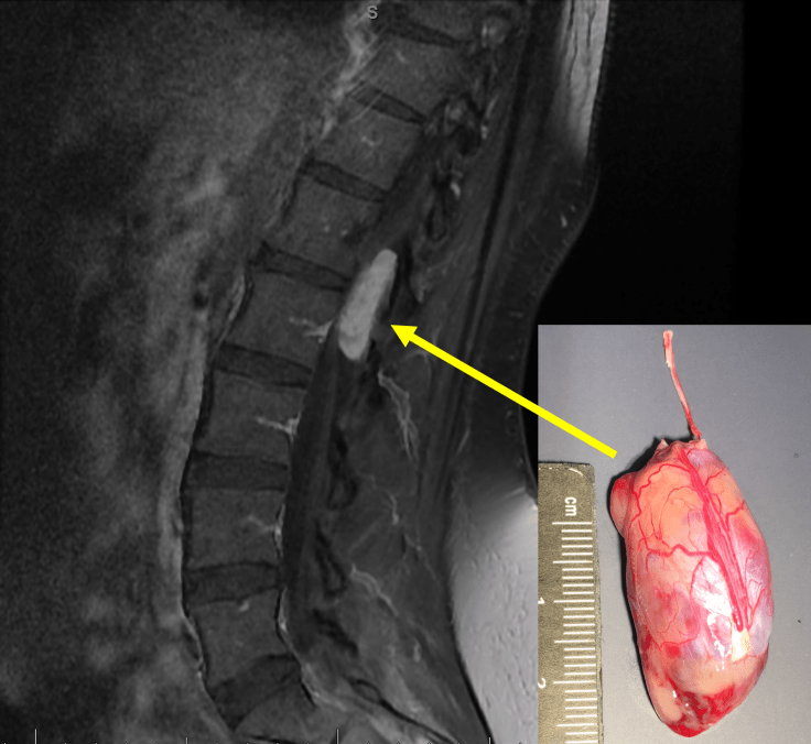
Myxopapillary ependymoma arises in the lumbar region of the spinal cord and typically produces symptoms associated with impingement of the spinal nerve roots of the cauda equina. It usually appears as an oval or sausage-shaped contrast-enhancing mass, like the one pictured in this MRI image (sagittal post-contrast T1 Fat-sat). The myxopapillary ependymoma in the inset photograph represents a gross surgical specimen received from a patient who had undergone optimal therapy: en bloc or gross total excision, in which the entire tumor is removed from the thecal sac without disturbance of the external capsule. Partial or incomplete tumor resection may result in spillage of tumor cells into the thecal sac and, consequently, these patients often experience local recurrence.
For further study on this topic and others related to neuropathology and neuroanatomy, the following books come highly recommended, and a small portion of the proceeds (or any Amazon purchase through these links) help with expenses of hosting this site:


Leave a comment