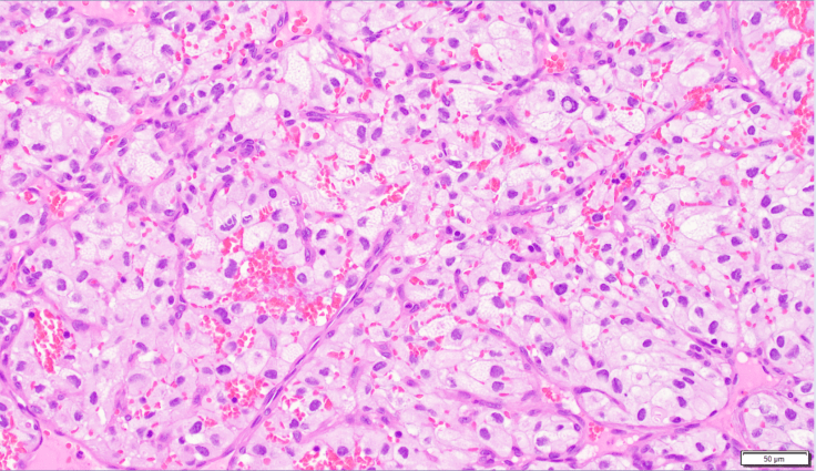
On gross examination, hemangioblastoma characteristically exhibits a yellow to yellow-orange color (similar to that of adipose tissue) due to the presence of lipid within the cytoplasm of neoplastic cells. On microscopic examination, the cells appear clear because the lipid that once filled the cytoplasm was washed out during tissue processing.
Hemangioblastoma usually arises within the cerebellum and represents one of the signature neoplasms commonly developed by persons with von Hippel Lindau (VHL) disease, which is, in addition to hemangioblastomas of the cerebellum as well as the retina, associated cysts of liver and pancreas, pheochromocytoma, and kidney tumors. The diagnosis that must be considered in the differential of hemangioblastoma, especially in VHL patients, is metastasis of the clear cell conventional subtype of Renal Cell Carcinoma (RCC). RCC is positive for Pax8 and CD10 immunostains, whereas hemangioblastoma is characteristically positive for Inhibin alpha.


Leave a comment